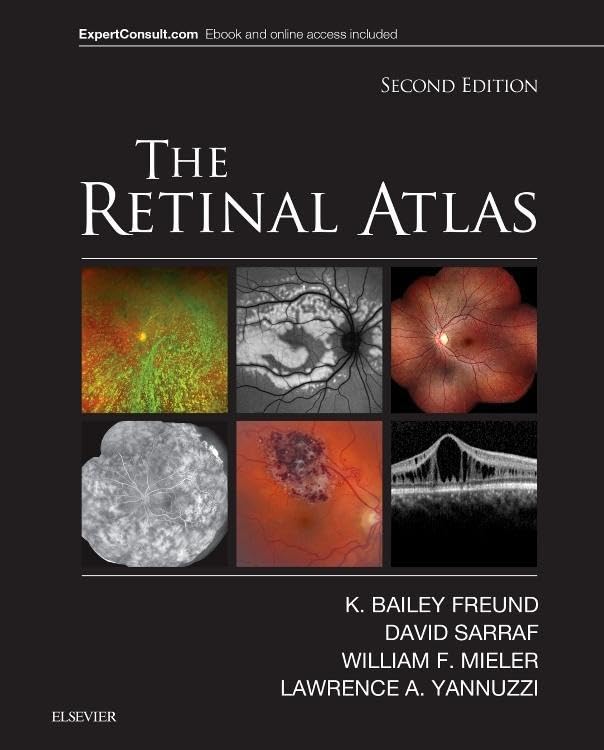
The Retinal Atlas, 2nd Edition
The Retinal Atlas is a second-edition clinical reference covering vitreous, retinal and macular disorders through extensive imaging and illustrations. Its detailed multimodal visual approach is aimed at ophthalmology professionals and trainees, rather than readers seeking a general eye-health guide.
At a glance
- Covers vitreous, retinal and macular disorders, including early and later disease stages.
- Includes examples across OCT, indocyanine green angiography, fluorescein angiography and fundus autofluorescence.
- Uses comparison images, colour coding, labels, arrows and magnified areas to draw attention to imaging findings.
- Describes spectral-domain and en face OCT, as well as ultra-wide-field imaging methods.
- Listing says an eBook version is included, with searchable text, figures and references across devices.
Often bought together
Pros and cons
Pros
- The broad disease coverage and multiple imaging methods make it relevant as a visual reference across retinal cases.
- Comparison layouts and annotated image details can help readers focus on lesion features and disease patterns.
- The listing states that the purchase includes a searchable eBook alongside the book.
Technical specifications
9 specifications| Edition | 2nd |
|---|---|
| Publisher | Elsevier |
| Publication date | 30 November 2016 |
| Language | English |
| Print length | 1200 pages |
| ISBN-10 | 0323287921 |
| ISBN-13 | 978-0323287920 |
| Weight listed | 215 g |
|---|---|
| Dimensions | 22.23 x 6.35 x 27.94 cm |
What to know before buying
- The listing describes more than 5,000 images, but does not specify a binding format. Confirm the edition and format offered if those details matter, and check how the eBook is accessed.
Who is it for?
A good fit for
- Retina specialists and comprehensive ophthalmologists wanting an image-led clinical reference.
- Ophthalmology residents and fellows studying retinal disease and imaging.
Less suited to
- Readers looking for a general introduction to eye health or a patient-focused guide.
- Buyers who need confirmation of a particular binding or eBook access method before purchasing.
Frequently asked questions
What subjects does The Retinal Atlas cover?
The listing describes coverage of vitreous, retinal and macular disorders, with images across several retinal imaging methods and disease stages.
Which imaging methods are included?
The listing names OCT, including spectral-domain and en face OCT, indocyanine green angiography, fluorescein angiography, fundus autofluorescence and ultra-wide-field imaging.
Is an eBook included?
The listing claims an eBook is included and describes searchable text, figures and references. Confirm the access method for the specific offer.
Who is this reference intended for?
The listing positions it for retina specialists, comprehensive ophthalmologists, residents and fellows.
Alternative options
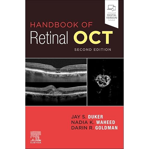
Handbook of Retinal OCT: Optical Coherence Tomography
$184.34
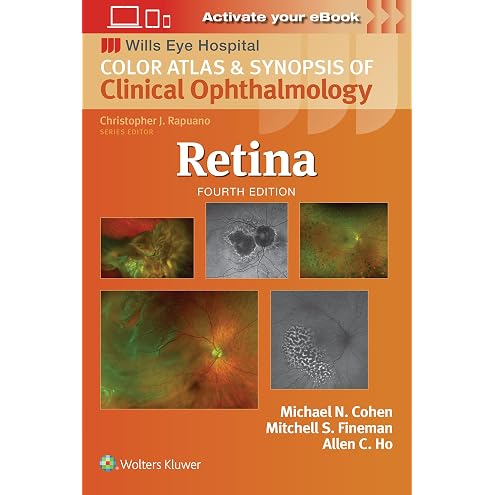
Color Atlas and Synopsis of Clinical Ophthalmology - Retina: Wills Eye Institute Atlas Series
$162.00
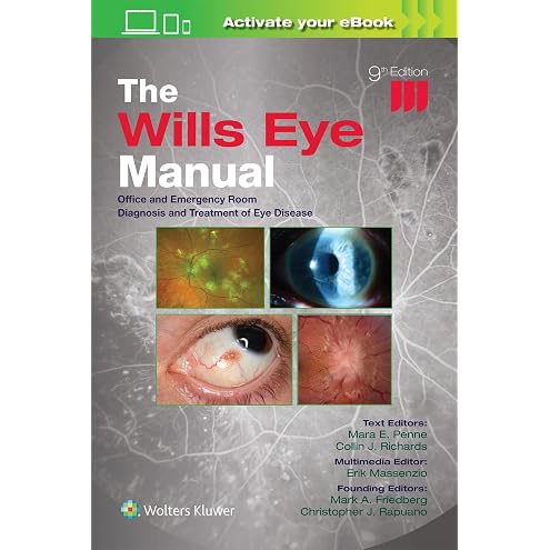
The Wills Eye Manual: Office and Emergency Room Diagnosis and Treatment of Eye Disease
$100.46

Portuguese: How to Get Really Good at Portuguese: Learn Portuguese to Fluency and Beyond
$13.19

Fluent Spanish through Short Stories
$15.99

Italian Short Stories for Beginners and Intermediate Learners: Learn Italian and Build Your Vocabulary (Italian Edition)
$4.99

Italian Conversation Made Natural: Engaging Dialogues to Learn Italian (Italian Edition)
$3.99
The Amazing 30-Day Guide to Drawing 3D Optical Illusions: How to Draw Spectacular Art That Leaps Off the Page With Easy Step-by-Step Guides
$20.31

French: How to Get Really Good at French: Learn French to Fluency and Beyond
$15.99